File:Giardia lamblia SEM 8698 lores.jpg
Jump to navigation
Jump to search

Size of this preview: 509 × 599 pixels. Other resolutions: 204 × 240 pixels | 626 × 737 pixels.


Original file (626 × 737 pixels, file size: 27 KB, MIME type: image/jpeg)

| Description |
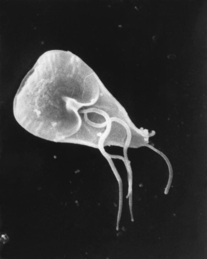
English: This scanning electron micrograph (SEM) revealed some of the external ultrastructural details displayed by a flagellated Giardia lamblia protozoan parasite. G. lamblia is the organism responsible for causing the diarrheal disease "giardiasis". Once an animal or person has been infected with this protozoan, the parasite lives in the intestine, and is passed in the stool. Because the parasite is protected by an outer shell, it can survive outside the body, and in the environment for long periods of time.
Cysts are resistant forms and are responsible for transmission of giardiasis. Both cysts and trophozoites can be found in the feces (diagnostic stages). The cysts are hardy and can survive several months in cold water. Infection occurs by the ingestion of cysts in contaminated water, food, or by the fecal-oral route (hands or fomites). In the small intestine, excystation releases trophozoites (each cyst produces two trophozoites). Trophozoites multiply by longitudinal binary fission, remaining in the lumen of the proximal small bowel where they can be free or attached to the mucosa by a ventral sucking disk. Encystation occurs as the parasites transit toward the colon. The cyst is the stage found most commonly in non-diarrheal feces. Because the cysts are infectious when passed in the stool or shortly afterward, person-to-person transmission is possible. While animals are infected with Giardia, their importance as a reservoir is unclear. |
| Source | http://phil.cdc.gov/PHIL_Images/8698/8698_lores.jpg |
| Author | CDC / Mahmud Tari |
| Permission (Reusing this file) |
Copyright Restrictions: None - This image is in the public domain and thus free of any copyright restrictions. As a matter of courtesy we request that the content provider be credited and notified in any public or private usage of this image. |

|
This file is a work of the Centers for Disease Control and Prevention, part of the United States Department of Health and Human Services, taken or made as part of an employee's official duties. As a work of the U.S. federal government, the file is in the public domain.
|
 |

|
This media comes from the Centers for Disease Control and Prevention's Public Health Image Library (PHIL), with identification number #8698. Note: Not all PHIL images are public domain; be sure to check copyright status and credit authors and content providers.
|
File history
Click on a date/time to view the file as it appeared at that time.
| Date/Time | Thumbnail | Dimensions | User | Comment | |
|---|---|---|---|---|---|
| current | 13:31, 6 July 2022 | | 626 × 737 (27 KB) | wikimediacommons>Chiswick Chap | crop deadspace |
File usage
The following page uses this file:
